CASE REPORT
Parotid Ductal Carcinoma with skin metastasis
Carcinoma Ductal Parotídeo con metástasis cutánea
Jadier
Wong-Silva1 ![]() *, Lucien
Gregoria Bory-Porras2
*, Lucien
Gregoria Bory-Porras2 ![]() ,
Mario Ramírez-Gómez3
,
Mario Ramírez-Gómez3 ![]()
1Universidad de Ciencias Médicas de Pinar del Río. Hospital Pediátrico Provincial Docente “Pepe Portilla”. Pinar del Río, Cuba.
2Universidad de Ciencias Médicas de Pinar del Río. Hospital General Docente “Abel Santamaría Cuadrado”. Pinar del Río, Cuba.
3Universidad Anáhuac México, Campus Norte. México
Cite as: Wong-Silva J, Bory-Porras LG, Ramírez-Gómez M. Parotid Ductal Carcinoma with skin metastasis. Odontologia (Montevideo). 2024; 2:133. https://doi.org/10.62486/agodonto2024133
Submitted: 12-02-2024 Revised: 15-05-2024 Accepted: 18-08-2024 Published: 19-08-2024
Editor:
Nairobi Hernández Bridón ![]()
ABSTRACT
Introduction: ductal carcinoma of the parotid salivary gland, have an epithelial origin. The behavior of this disease is aggressive, so a correct and early diagnosis is vital. In this research a case with a diagnosis of parotid ductal carcinoma with metastatic skin lesions is presented.
Case presentation: 60-year-old male patient with a history of high blood pressure who for approximately 3 months has been experiencing discomfort in the pre-auricular region, lower third of the right side of the face, and exophytic lesions in the skin of that area. An oral examination revealed poor oral hygiene, the presence of root remains, an increase in the volume of the excretory duct of the right parotid gland, and changes in the integrity of the buccal mucosa. Complementary and imaging tests are indicated (imaging, biopsy, computed axial tomography and immunohistochemistry). The diagnosis was confirmed with a T4N2M1 staging and total surgical treatment was determined immediately.
Conclusions: patients with ductal carcinoma, despite treatment with parotidectomy, cervical dissection and postoperative radiotherapy, a large proportion of patients develop local and distant recurrence, so a timely diagnosis can guarantee greater survival for the patient.
Keywords: Parotid; Cancer; Oncogenes; Biopsy.
RESUMEN
Introducción: el carcinoma ductal de la glándula salival parótida, tiene un origen epitelial y poco frecuente. El comportamiento de esta patología es agresivo por lo que un correcto y temprano diagnóstico es vital. En esta investigación se presenta un caso con diagnóstico de carcinoma ductal parotídeo con lesiones metastáticas cutáneas.
Presentación de caso: paciente masculino de 60 años de edad con antecedentes de hipertensión arterial que hace aproximadamente 3 meses presenta molestias en la región prearicular, tercio inferior de la hemicara derecha y lesiones exofíticas en la piel de esa zona. Al examen bucal se observa deficiente higiene bucal, presencia de restos radiculares, aumento de volumen del conducto excretor de la glándula parótida derecha y cambios en la integridad de la mucosa yugal. Se indican exámenes complementarios y de imagen (imagenológicos, biopsia, tomografía axial computarizada e inmunohistoquímica). Fue confirmado el diagnóstico con una estadificación T4N2M1 y se determina tratamiento quirúrgico total de inmediato.
Conclusiones: los pacientes con carcinoma ductal a pesar del tratamiento con parotidectomía, vaciamiento cervical y radioterapia postoperatoria, una gran proporción de pacientes desarrollan recurrencia local y a distancia, por lo que un diagnóstico oportuno puede garantizar una mayor sobrevida para el paciente.
Palabras clave: Parótida; Cáncer; Oncogenes; Biopsia.
INTRODUCTION
Ductal carcinoma of the salivary glands is a rare tumor, with an incidence of 1 % to 3 % of all malignant epithelial neoplasms of the salivary gland. In 88 % of cases, it is located in the parotid gland and accounts for 0,9-6 % of all parotid tumors. It most frequently affects men, with a peak incidence between the seventh and eighth decade of life, coinciding in age, sex, and location with the case we describe.(1)
Clinically, it presents as a painful parotid tumor associated with facial paralysis in 40-60 % of cases, with lymph node involvement in 40-80 % and distant metastasis in 35-65 % of patients.(2) Radiologically, magnetic resonance imaging (MRI) is the test of choice for studying these tumors' limits and degree of infiltration, with a sensitivity of 80 % and 89 %, respectively. However, CT is essential for the evaluation of bone involvement. Histologically, it presents similar characteristics to ductal breast carcinoma, so it is important to rule out mammary gland metastasis. It has an intraductal and an infiltrative carcinomatous component. The intraductal component presents different growth patterns, usually cribriform, papillary, solid, and comedocarcinoma type, and the infiltrative component is a desmoplastic stroma.(1,2,3)
Immunohistochemically, overexpression of cytokeratin (CAM5.2), cytokeratin (AE1/AE), cytokeratin, HMW-CK 34ßE12 (high-molecular-weight cytokeratin), EMA (epithelial membrane antigen), GCDFP-15 (gross cystic disease fluid protein-15), AR (androgen receptor) and HER2/neu has been identified in ductal carcinoma of salivary glands. The overexpression of the c-erbB-2 protein is associated with a worse prognosis. Based on this, some studies suggest that hormonal therapy with Trastuzumab (a monoclonal antibody that blocks the extracellular domain of the HER2/neu receptor) increases the survival rate.(4,5)
Treatment should be multidisciplinary, based on surgery (total parotidectomy and cervical emptying), radiotherapy, and chemotherapy, in order to achieve better control of recurrences and distant metastases. The prognosis of ductal carcinoma of salivary glands is unfavorable, with a local recurrence rate of 33 %, lymph node metastases of 59 %, distant metastases of 46 %, and a 4-year mortality rate of 65 %.(6)
Parotid ductal carcinoma is a poor prognostic tumor. The local recurrence rate is estimated at 13-48 % (3,4), lymph node involvement at around 40-80 %,(4,8) and distant metastases are detected in 35-65 % of cases.(1,6)
Numerous factors associated with poor prognosis have been described in the literature. Among them, we highlight male sex, high tumor size (T), affected lymph nodes (N+), advanced age, positive surgical margins, and perineural and lymphovascular invasion. Survival is generally around five years for 40 to 60 % of diagnosed cases. (16, 17)
CASE REPORT
A 60-year-old male patient with a history of arterial hypertension (AHT) who approximately three months ago presented with discomfort in the preauricular region and lower third of the right hemiface. It was interpreted and diagnosed as herpes zoster. The corresponding treatment was indicated, and due to the unfavorable course of the symptoms, he was referred to the Maxillofacial Surgery Service, where he was examined and evaluated.
General physical examination showed an increased volume in the preauricular region and lower third of the right hemiface (figure 1), with signs of facial paralysis and swelling in the area of the right parotid gland, of firm consistency, painful on palpation; cervical adenopathies were detected.
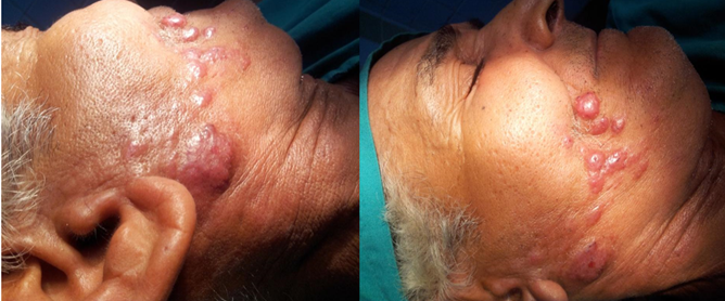
Figure 1. Presence of volume increase and facial lesions
The oral examination revealed poor oral hygiene, presence of root debris, increased volume of the excretory duct of the right parotid gland and changes in the integrity of the jugal mucosa (Figure 2).
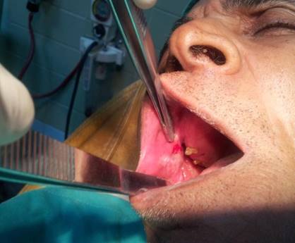
Figure 2. Oral examination
Complementary and imaging tests (imaging, biopsy, computed axial tomography, and immunohistochemistry) are indicated.
Complementary Examinations
· Platelet count 300 x 109/L
· Lymph: 5,3 x 10⁹/L,
· Lymph: 29 %
· PMN: 60 %
· Erythro: 5 mm/h
· Blood glucose: 4,7 mmol/L
· TGP: 14 U/I
· TGO: 24 U/I
· Alkaline phosphatase: 132 U/I.
· Total proteins: 70 g/l
· Total bilirubin: 5
· Creatinine: 90 mmol/l
· LDH: 134 mmol/l
· HCV, HBV, HIV, VDRL : Negative
Imaging, anatomopathological and immunohistochemical studies were indicated. In ultrasound a simple imaging study was performed, where the gland involved was found to be heterogeneous, with irregular edges and hyperdense and thickened excretory duct, with lesions at the expense of its wall.
Computed Axial Tomography (contrasted) (Neck and Thorax) showed a nodular volume increases in the right parotid region, irregular borders, and involvement of the VII cranial nerve without bone involvement, which is to be defined by PET-CT.
A fine needle aspiration biopsy was performed in agreement with the initial ultrasound, and it showed epithelial neoplasm (oncocytic) without lymphoid content. The neoplastic cells showed positivity for GCDFP-15 and CK34betaE12 markers and negativity for Galectin-3, S100, and P63. Perineural infiltration, vascular infiltration, and bone infiltration in the right hemimandible were observed. The studies performed up to that moment allowed us to define that the patient presented a parotid ductal carcinoma.
It was determined to perform PET-CT, which confirmed the presence of locoregional adenopathies < 6cm and the capture of the posterior border of the right mandibular ramus, although the presence of distant metastasis was ruled out. Tumor staging was T4N2M1.
Right total parotidectomy with right hemimandibulectomy and cervical lymph node removal was indicated. In addition, as a supportive and complementary treatment, radiotherapy was given.
DISCUSSION
Ductal carcinoma of the salivary glands is a very rare adenocarcinoma generated from the cells of the excretory gland duct. The published incidence varies from 3-9 % of malignant salivary gland tumors. However, it is difficult to determine the true incidence rate of this tumor since it has not been recognized in numerous series or has been confused with other tumors due to the little knowledge that exists about it. (1,7)
They usually arise de novo, although there are cases developed from a pleomorphic adenoma and less frequently from a low-grade adenocarcinoma. It manifests as a painful mass frequently associated with facial nerve paresis/paralysis.
Cases have been described in young patients and locations other than the salivary glands, such as the larynx and paranasal sinuses. However, its most common presentation is in males between 50 and 60 years of age with parotid gland involvement. CT findings of tumor calcifications may suggest CDS in 33 % of cases. MRI helps to evaluate signs suggestive of malignancy, such as poorly defined margins and comedonecrosis. It is advisable to perform PET-CT to diagnose and follow up on possible distant metastases. (8,9)
The main cause of death is distant metastases in the lung and bone, although cases have been described with survival of more than ten years, in which it can be said that there is a criterion of cure for the disease. There is controversy as to the factors of poor prognosis, although tumor size and lymph node involvement stand out, followed by distant metastases and Her-2/neu expression. (10,11)
Histologically, CDS is characterized by its great similarity to ductal breast carcinoma, with areas of invasion and areas of intraductal carcinoma. The intraductal pattern presents different types of growth, such as cribriform, papillary, and solid, generally associated with areas of central comedonecrosis. (12) The invasive pattern presents a desmoplastic stroma; immunohistochemically, it presents strong positivity for RA and cytokines. (13,14)
As for treatment, total parotidectomy with ipsilateral cervical emptying, radiotherapy, and chemotherapy are recommended. In advanced cases with distant metastases, some studies show favorable results after treatment with trastuzumab, a monoclonal antibody selective for Her-2/neu, expressed in ductal breast carcinoma. (15)
The ideal test to complete the extension study is PET-CT, which will show a highly metabolic tumor in the parotid area and can locate regional and distant metastases. (16)
We will need histopathological confirmation with the characteristics described above to diagnose parotid ductal carcinoma. The sample can be obtained by fine needle puncture-aspiration, but we will need to perform an excision of the sample through surgery for a definitive diagnosis.
Given the aggressive characteristics of this tumor, the treatment of choice is radical parotidectomy (with or without preservation of the facial nerve) associated with ipsilateral cervical lymph node emptying and radiotherapy. (17) Radiotherapy is indicated in the presence of histopathological features of poor prognosis, such as positive surgical margins, lymphatic or perineural invasion, and extra parotid extension. (12)
CONCLUSIONS
Parotid ductal carcinoma is a malignant tumor with aggressive characteristics, and in most patients, it is diagnosed in advanced stages. It has specific histopathological characteristics; in patients with ductal carcinoma, despite treatment with parotidectomy, cervical emptying, and postoperative radiotherapy, a large proportion of patients develop local and distant recurrence so that a timely diagnosis can ensure greater survival for the patient.
REFERENCES
1. ANDREADIS D et al. Detection of C-KIT (CD117) molecule in benign and malignant salivary gland tumours. Oral Oncol [Internet] 2006 [citado 03/05/2024]; 42 (1): 57-65. Disponible en: https://pubmed.ncbi.nlm.nih.gov/16140564/
2. EVESON JW et al. Tumours of the salivary glands. In: LEON BARNES, L. et al. (Ed.) Pathology and genetics. Head and neck tumours. World Health Organization Classification of Tumours. Lyon: IARCPress [Internet] 2008 [citado 03/05/2024]; 244. Disponible en: https://publications.iarc.fr/Book-And-Report-Series/Who-Classification-Of-Tumours/Pathology-And-Genetics-Of-Head-And-Neck-Tumours-2005
3. FANG X et al. Osteoclast-like giant cell tumor of the salivary gland. Ann Diagn Pathol [Internet] 2009 [citado 03/05/2024]; 13 (2): 114-8. Disponible en: https://pubmed.ncbi.nlm.nih.gov/19302960/
4. Karaman E, Saritzali G, Kilic E, Korkut N, Enver O. Follicular dendritic cell sarcoma of the parotid gland recurring 6 times within 12 years. J Craniofac Surg. [Internet] 2009 [citado 03/05/2024]; 20(6):2171-2. Disponible en: https://pubmed.ncbi.nlm.nih.gov/19884836/
5. Liess BD, Hirschi S, Zitsch RP 3rd, Frazier S, Konrad A. Carcinosarcoma of the parotid gland: report of a case with immunohistochemical findings. Ann Otol Rhinol Laryngol. [Internet] 2007 [citado 03/05/2024]; 116(9):702-4. Disponible en: doi: 10.1177/000348940711600913. PMID: 17926594.
6. QURESHI, A. True malignant mixed tumor of parotid. J Coll Physicians Surg Pak [Internet] 2007 [citado 03/05/2024]; 17 (11): 697-8. Disponible en: https://pubmed.ncbi.nlm.nih.gov/18070582/
7. Staffieri C, Marioni G, Ferraro SM, Marino F, Staffieri A. Carcinosarcoma de novo of the parotid gland. Oral Surg Oral Med Oral Pathol Oral Radiol Endod. [Internet] 2007 [citado 03/05/2024]; 104(2):e35-40. Disponible en: https://pubmed.ncbi.nlm.nih.gov/17630097/
8. Vékony H, Leemans CR, Ylstra B, Meijer GA, van der Waal I, Bloemena E. Salivary gland carcinosarcoma: oligonucleotide array CGH reveals similar genomic profiles in epithelial and mesenchymal components. Oral Oncol [Internet] 2009 [citado 03/05/2024]; 45(3):259-65. Disponible en: doi: 10.1016/j.oraloncology.2008.05.009. Epub 2008 Aug 6. PMID: 18693132.
9. Granell JL, Sánchez-Jara J, Gavilanes MJ Velasco, T Collazo, J Herrero, et al. Manejo de la enfermedad quirúrgica de la glándula parótida: revisión de 54 casos Acta Otorrinolaringol Esp [Internet] 2010 [citado 03/05/2024]; 61: 189–195. Disponible en: https://pesquisa.bvsalud.org/portal/resource/pt/ibc-87756
10. Wang WH, Wang CY, Bian L, Xia B, Hu YY, Xu B. Study about the clinical and pathological characteristics of salivary duct carcinoma. Hua Xi Kou Yi Xue Za Zhi. [Internet] 2010 [citado 03/05/2024]; 28:128-131. Disponible en: http://www.ncbi.nlm.nih.gov/pubmed/20480651
11. Kaidar-Person O, Billan S, Kuten A. Targeted therapy with trastuzumab for advanced salivary ductal carcinoma: case report and literatura review Med Oncol [Internet] 2011 [citado 03/05/2024]. Disponible en: https://www.springerlink.com/content/n04h473260149608/
12. General Medical Council. Workplace Based Assessment: A Guide for Implementation. A GMC/AoMRC Guidance Paper [Internet] 2010 [citado 03/05/2024]. Available at: http://www.gmc-uk.org/Workplace_Based_Assessment-A_guide_for_implementation_0410.pdf_48905168.pdf.
13. Curriculum for the Foundation Years in Postgraduate Education and Training Foundation Programme Committee of the Academy of the Royal Colleges, in cooperation with Modernising Medical Careers in the Departments of Health. [Internet] 2013 [citado 03/05/2024]. Available at http://www.mmc.nhs.uk/pages/foundation/Curriculum 2005.
14. Mlika M, Kourda N, Zidi Y, Aloui R, Zneidi N,Rammeh S, Zermani R and Jilani S. Salivay duct carcinoma of the parotid gland. J Oral Maxillofac Pathol. [Internet] 2012 [citado 03/05/2024]; 16(1):134-6. Disponible en: https://www.ncbi.nlm.nih.gov/pmc/articles/PMC3303509/
15. Kristensen RN, Hahn CH. Facial nerve palsy caused by parotid gland abscess. J Laryngol Otol [Internet] 2012 [citado 03/05/2024]; 126: 322-4. Disponible en: https://pubmed.ncbi.nlm.nih.gov/22017803/
16. D’Heygere E, Meulemans J, Vander Poorten V. Salivary duct carcinoma. Curr Opin Otolaryngol Head Neck Surg. [Internet] 2018 [citado 03/05/2024]; 26(2):142-151. Disponible en: https://pubmed.ncbi.nlm.nih.gov/29373327/
17. Nijim-Nijim H, Díez-González L, Elhendi-Halawa W. Adenocarcinoma ductal de parótida. An Orl Mex. [Internet] 2015 [citado 03/05/2024]; 60:261-264. Disponible en: https://www.revista-portalesmedicos.com/revista-medica/carcinoma-ductal-de-parotida-a-proposito-de-un-caso-clinico/
CONFLICTS OF INTEREST
The authors declare that there is no conflict of interest.
FUNDING
The authors did not receive funding for the development of this research.
AUTHORSHIP CONTRIBUTION
Conceptualization: Jadier Wong-Silva, Lucien Gregoria Bory-Porras, Mario Ramírez-Gómez.
Formal analysis: Jadier Wong-Silva, Lucien Gregoria Bory-Porras, Mario Ramírez-Gómez.
Writing - initial draft: Jadier Wong-Silva, Lucien Gregoria Bory-Porras, Mario Ramírez-Gómez.
Writing - revision and editing: Jadier Wong-Silva, Lucien Gregoria Bory-Porras, Mario Ramírez-Gómez.