CASE REPORT
Pleomorphic adenoma in an adolescent, about a clinical case
Adenoma pleomorfo en una adolescente, a propósito de un caso clínico
Damian
Soto-Castillo1 ![]() ,
Jadier Wong-Silva2
,
Jadier Wong-Silva2 ![]() *, Lucien
Gregoria Bory-Porras3
*, Lucien
Gregoria Bory-Porras3 ![]() ,
Mario Ramírez-Gómez4
,
Mario Ramírez-Gómez4 ![]()
1Hospital Pediátrico Provincial “Mártires de Las Tunas”. Las Tunas, Cuba.
2Universidad de Ciencias Médicas de Pinar del Río. Hospital Pediátrico Provincial Docente “Pepe Portilla”. Pinar del Río, Cuba.
3Universidad de Ciencias Médicas de Pinar del Río. Hospital General “Abel Santamaría Cuadrado”. Pinar del Río, Cuba.
4Universidad Anáhuac México, Campus Norte. México.
Cite as: Soto-Castillo D, Wong-Silva J, Bory-Porras LG, Ramírez-Gómez M. Pleomorphic adenoma in an adolescent, about a clinical case. Odontologia (Montevideo). 2024; 2:145. https://doi.org/10.62486/agodonto2024145
Submitted: 17-02-2024 Revised: 20-05-2024 Accepted: 19-08-2024 Published: 20-08-2024
Editor: Nairobi
Hernández Bridón ![]()
ABSTRACT
Introduction: salivary gland tumors in children represent a low percentage of pediatric head and neck tumors; most of them are diagnosed in association with the Parotid salivary gland and present a higher proportion of malignancy than in adults.
Objective: to present a case with a diagnosis of Pleomorphic Adenoma: clinical symptoms, complementary examinations, treatment and evolution.
Case presentation: female patient, 13 years old, from a rural background and white race, attended the Maxillofacial Surgery consultation, in good general condition, accompanied by her parents, due to an increase in volume of a hard consistency, not fixed to deep planes ( mobile), asymptomatic, of 7 months of evolution and 3 cm in length located in the left parotid region. Ultrasound of soft tissues and neck was indicated, as well as contrast-enhanced facial mass tomography, which showed a mass in relation to the left parotid gland that captured contrast. In addition, complementary examinations and a fine needle aspiration biopsy (FNAB) were performed, resulting in a benign parotid gland tumor (Pleomorphic Adenoma). Superficial parotidectomy was performed with preservation of the facial nerve. The patient progressed satisfactorily and continues to be followed up by consultation.
Conclusions: pleomorphic adenomas are discovered in practice as slowly growing asymptomatic masses; the size is variable and will always depend on the stage at which it is detected.
Keywords: Benign Tumor; Parotid Gland; Pleomorphic Adenoma.
RESUMEN
Introducción: los tumores de glándulas salivares en niños constituyen un bajo porciento de los tumores pediátricos de cabeza y cuello, de ellos, la mayoría son diagnosticados asociados a la glándula salival parótida y presentan una mayor proporción de malignidad que en los adultos.
Objetivo: presentar un caso con diagnóstico de Adenoma pleomorfo: sintomatología clínica, exámenes complementarios, tratamiento y evolución.
Presentación de caso: paciente de sexo femenino, 13 años de edad, procedencia rural y raza blanca, acudió a consulta de Cirugía Maxilofacial, con buen estado general acompañada por sus padres, por aumento de volumen de consistencia dura, no fijo a planos profundos (móvil), asintomático, de 7 meses de evolución y 3 cm de longitud localizado en región de parótida izquierda. Se indicó ultrasonido de tejidos blandos y cuello, tomografía de macizo facial contrastada la cual aportó una masa en relación con la glándula parótida izquierda que capto contraste. Además, le fueron realizados exámenes complementarios y una biopsia por aspiración con aguja fina (BAAF) dando como resultado un tumor benigno de glándula parótida (Adenoma Pleomorfo). Se llevó a cabo la parotidectomía superficial con conservación del nervio facial. La paciente evolucionó satisfactoriamente y sigue en seguimiento por consulta.
Conclusiones: los adenomas pleomorfos son descubiertos en la práctica como masas asintomáticas de lento crecimiento, el tamaño es variable, siempre dependerá de la etapa en la que sea detectado.
Palabras clave: Tumor Benigno; Glándula Parótida; Adenoma Pleomorfo.
INTRODUCTION
Salivary gland tumors are relatively uncommon and usually manifest as swelling in one of the paired major salivary glands or in one of the minor glands of the mouth, accounting for about 3 to 6 % of all head and neck tumors. The incidence of these tumors is increasing concerning lesions of the epithelial lineage of the upper aerodigestive tract.(1,2)
In children, they account for less than 10 % of pediatric head and neck tumors. They are malignant in a high percentage, which, according to the series, is estimated to be as high as 50 % if vascular malformations are excluded. They affect 80 % of the parotid gland and present a higher proportion of malignancy than in adults. (3)
Pleomorphic adenoma or mixed tumor is the most common of all salivary gland tumors and represents approximately 70 % of all parotid gland tumors. These tumors have a female predilection and are most commonly seen between the third and sixth decades of life. They are usually discovered during routine physical examination as an enlargement of glandular origin in the head and neck region that does not cause ulceration of the overlying skin and mucosa. (4, 5)
Pleomorphic adenomas are discovered in practice as asymptomatic masses of slow growth, and their size is variable, always depending on the stage at which they are detected. Among the symptoms described by patients are Foreign body sensation, otalgia, trismus, pain, and facial paralysis. (11)
Surgical resection, partial surgery, and subtotal or total parotidectomy with preservation of the facial nerve are usually the indicated treatments. Pleomorphic adenoma is a benign tumor that may recur or recur and may also present malignant transformation. The prognosis is generally favorable, with a 95 % cure rate. (12)
We present below a case with a diagnosis of pleomorphic adenoma in the parotid gland, resolved surgically by superficial parotidectomy.
CASE REPORT
We present a case of a 13-year-old female patient of rural origin and a white race with a medical history. She came to the Maxillofacial Surgery office in good general condition, accompanied by her parents, due to an increase in the volume of hard consistency, not fixed to deep planes (mobile), asymptomatic, of 7 months of evolution and 3 cm in length, located in the left parotid region.
Ultrasound of soft tissues and neck and contrasted tomography of facial mass was indicated, which showed a mass about the left parotid gland that captured contrast, defined, and encapsulated. In addition, complementary examinations and a fine needle aspiration biopsy (FNA) were performed, resulting in a benign tumor of the parotid gland (pleomorphic adenoma).
After a thorough analysis of all the results of the examinations and with the consent of the parents, it was decided to operate on the patient, and a superficial parotidectomy with preservation of the facial nerve was performed. The sample was sent to the Anatomic Pathology department for histopathology study, giving a definitive result: a pleomorphic adenoma. The patient evolved satisfactorily and continues to be followed up in consultation.
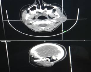
Figure 1. Contrast tomography image of the facial mass showing a mass in relation to the left parotid gland
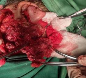
Figure 2. Superficial parotidectomy for pleomorphic adenoma with preservation of the facial nerve.
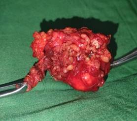
Figure 3. Parotid tumor (pleomorphic adenoma)
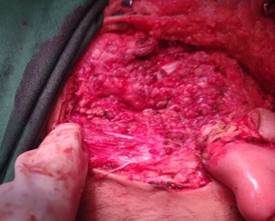
Figure 3. Facial nerve preserved after surgery
DISCUSSION
Pleomorphic adenomas are generally solitary; however, they can be synchronously or metachronously associated with other tumors, particularly with Warthin's tumor, either in the same gland or different glands.(6)
Generally, pleomorphic adenomas arise as asymptomatic masses of slow growth, varying from a few millimeters to several centimeters, reaching considerable sizes in some cases. In addition, more symptoms such as foreign body sensation, otalgia, parotid mass, and trismus are described.(7)
In the inspection, the existence or not of asymmetries must be objectified. Parotid palpation compares the size and consistency of the two glands and ends with bimanual or digital palpation by placing a hand inside the oral cavity. By expressing Stenon's or Wharton's ducts, it is possible to observe the characteristics of the saliva produced by the various glands. If an inflammatory disease is excluded, pain on palpation of the parotid gland is usually a sign of malignancy. The examination is completed with an inspection of the oral cavity. (8)
Histologically, they show mixed epithelial and mesenchymal differentiation, with scattered epithelial nests in a variable matrix with hyaloid, chondroid, or osseous myxoid differentiation. Once the initial diagnosis is made, the treatment of pleomorphic adenoma is purely surgical; in fact, it is considered the mainstay in the therapeutic management of this pathology. (9)
Superficial parotidectomy with preservation of the facial nerve is the most commonly indicated procedure; in addition, submandibular sialoadenectomy or wide local excision for a minor salivary gland can be performed. The common point of all these modalities is the need to perform a meticulous capsule dissection to avoid neoplasm recurrence. (8)
As for the malignant transformation of pleomorphic adenoma in general, it is reported in 1.5 % to 23 % of the cases, and it is described that this increases according to the time of evolution of the disease since carcinogenesis occurs in several stages through genomic changes that result in the loss of functions of tumor suppressors, activation of oncogenes and fusion of genes with malignant potential.(10)
CONCLUSIONS
Among the main complications of surgical resection of pleomorphic adenoma are hemorrhage, sialocele, and facial paralysis. The latter accounts for about 15 % of the complications, although usually temporary. Facial paralysis can be prevented by detailed dissection of the facial nerve.
REFERENCES
1. Hernando M, Martín Fragueiro L, Eisenberg G, Echarri R, García Peces V, Urbasos M, et al. Tratamiento quirúrgico de los tumores de glándulas salivales. Acta Otorrinolaringol Esp [Internet] 2009 [citado 03/05/2024]. Disponible en: https://www.clinicalkey.es//#!/content/journal/1- s2.0S0001651909000491
2. Rivero Granado CE, León Céspedes IM, Vázquez Blanco E, Valerino Guzmán E, Vázquez Ortiz HJ, Caracterización de pacientes con neoplasias malignas de la glándula parótida atendidos en el hospital “Celia Sánchez Manduley. Revista Electrónica Dr. Zoillo E. Marinello Vidaurreta [Internet] 2022 [citado 03/05/2024]. Disponible en: http://revzoilomarinello.sld.cu/index.php/zmv/article/view/3085.
3. M.S. Fernández Córdoba, J. González Piñera, J.P. García De La Torre, O. Sánchez París, R Parrado Villodres, M. Lillo Lillo. Tumores de parótida en niños. Revista Cirugía Pediátrica [Internet] 2008 [citado 03/05/2024]; 21 (1). Disponible en: http://www.secipe.org/coldata/upload/revista/20080107.pdf
4. Díaz Pérez, Carlos Alberto; Simons Preval, Sara Jane; Martínez Rodríguez, Milagros ADENOMA PLEOMÓRFICO EN EDAD PEDIÁTRICA. PRESENTACIÓN DE 3 CASOS Revista Información Científica [Internet] 2011 [citado 03/05/2024]; 72 (4). Disponible en: http://www.redalyc.org/articulo.oa?id=551757294029
5. Hernández García DG, Fernández Estrada XI, Lagunes López MA, Rivera Macías Rodríguez Tomás JA, Camacho Olguín CG.Adenoma pleomorfo en glándula parótida: reporte de un caso. Ciencia en la frontera: revista de Ciencia y Tecnología de la UACJ [Internet] 2019 [citado 03/05/2024]; 15 (1). Disponible en: http://erevistas.uacj.mx/ojs/index.php/cienciafrontera/article/view/3920
6. Morales GM, Durán EE, Fonseca MG, Valente B. Eficacia de la biopsia por aspiración con aguja fina en enfermedad de las glándulas salivales. An Orl Mex [Internet] 2020 [citado 03/05/2024]. Disponible en: https://www.medigraphic.com/pdfs/anaotomex/aom-2020/aom201c.pdf
7. Reis A, Miranda M, Koch T, Morosolli A. Diagnóstico ecográfico do adenoma pleomórfico da parótida: caso clínico. Revportes Tomatol Med Cirmaxilofac [Internet] 2016 [citado 03/05/2024]. Disponible en: http://dx.doi.org/10.1016/j.rpemd.2016.
8. LBauta-Milord R, Góngora-Gómez O, Gómez-Vázquez YE. Caracterización clínica y anatomopatológica del adenoma pleomórfico de glándulas salivales. Univ Méd Pinareña [Internet] 2020 [citado 03/05/2024]: e 519. Disponible en: http://www.revgaleno.sld.cu/index.php/ump/article/view/519
9. Barragán W, Cueva J, Cedeño M, Regalado R, Villacrés D. Adenoma Pleomorfo de Parótida, Manejo Quirúrgico: A Propósito de un caso. Rev Med HJCA [Internet] 2020 [citado 03/05/2024]. http://dx.doi.org/10.14410/2020.12.1.cc.10
10. A Abarca, D Peñaloza, A Urrutia, M Cancino. ADENOMA PLEOMORFO ORIGINADO EN PARED LATERAL NASAL: REPORTE DE UN CASO CLÍNICO. Revista de Otorrinolaringología y Cirugía de Cabeza y Cuello [Internet] 2019 [citado 03/05/2024]. Disponible en: http://www.scielo.cl/scielo.php?script=sci_arttext&pid=S0718-48162019000300341
11. Bahbah S, Chbicheb S. Pleomorphic adenoma of the cheek. Case report with review. Int. J. Odontostomat [Internet] 2020 [citado 03/05/2024]; 14(4):653-657. Disponible en: http://www.ijodontostomatology.com/wp-content/uploads/2020/08/2020_v14n4_034.pdf
12. Avigeet G, Koochakzadeh S, Neskey D, Nguyen S, Carcinoma ex pleomorphic adenoma: A review of incidence, demographics, risk factors, and survival. Am J. Otolaryngol [Internet] 2019 [citado 03/05/2024]; 40(6). DOI: https://doi.org/10.1016/j.amjoto.2019.102279
FINANCING
The authors did not receive funding for the development of this research.
CONFLICT OF INTEREST
The authors declare that there is no conflict of interest.
AUTHORSHIP CONTRIBUTION
Conceptualization: Damian Soto-Castillo, Jadier Wong-Silva, Lucien Gregoria Bory-Porras, Mario Ramírez-Gómez.
Formal analysis: Damian Soto-Castillo, Jadier Wong-Silva, Lucien Gregoria Bory-Porras, Mario Ramírez-Gómez.
Writing - initial draft: Damian Soto-Castillo, Jadier Wong-Silva, Lucien Gregoria Bory-Porras, Mario Ramírez-Gómez.
Writing - revision and editing: Damian Soto-Castillo, Jadier Wong-Silva, Lucien Gregoria Bory-Porras, Mario Ramírez-Gómez.